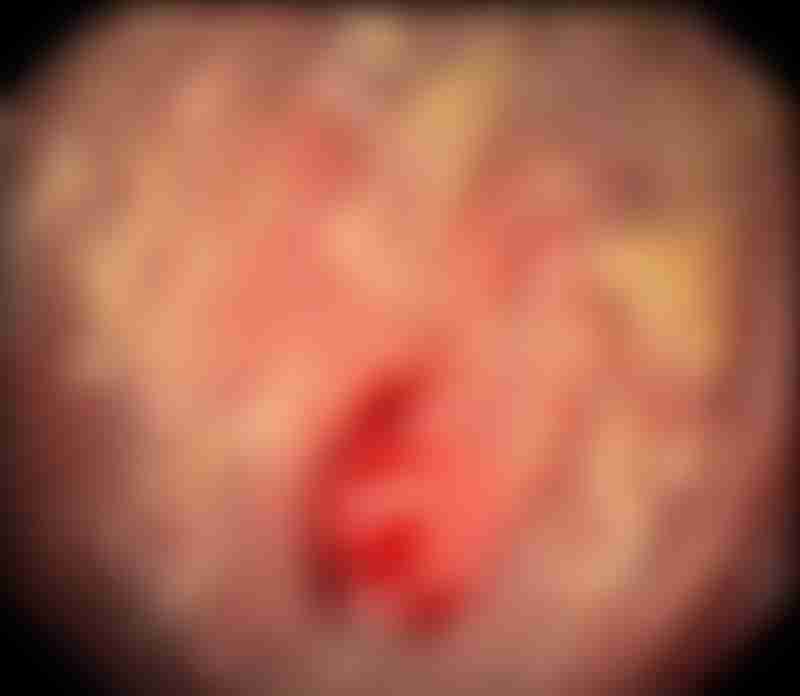
Types and Grades of Gastric Neuroendocrine Tumors

Source: Shutterstock
What is a gastric neuroendocrine tumor?
Neuroendocrine tumors (NETs) are rare cancers that can occur anywhere in the body. Those that originate from neuroendocrine cells located in the mucosal lining of the stomach are called gastric NETs. These specialized cells produce hormones controlling the release of gastric juices and how quickly food moves through the stomach.

Light micrograph of tissue from a gastric neuroendocrine tumor (purple mass at center). Source: Cnri/Science Photo Library
Gastric NETs can be classified according to their types and grades based on information that doctors have gathered from diagnostic scans and laboratory tests. Understanding the different types and grades is essential for patients as it affects the treatment plan and prognosis. Patients can use this information to make informed decisions about their treatment and to communicate effectively with their doctors.
Types of gastric neuroendocrine tumors
There are three types of gastric NETs.
Type I
Type I gastric NETs are the most common type, accounting for 70% to 80% of all NETs originating from the stomach. These tumors are often small (less than 1 to 2 cm long), but many. They occur mostly in the gastric body and fundus, and are usually limited to the gastric mucosa and submucosa. Type I gastric NETs are also well-differentiated (appear similar to normal, healthy cells), rarely aggressive and unlikely to spread to other tissues and organs.

Endoscopic view of a neuroendocrine tumor and atrophic gastritis in the stomach lining. Source: Gastrolab/Science Photo Library
Type I gastric NETs are often associated with a condition called atrophic gastritis, which causes the stomach cells to become irritated and inflamed. If not addressed, this can lead to issues with the production of stomach acid. These tumors also produce high levels of the hormone gastrin, which is a known proliferative stimuli of enterochromaffin cells (ECLs) that make up a gastric NET. The excess of gastrin therefore leads to hypertrophy and hyperplasia of the ECLs.
Type II
This type of gastric NET is a lot less common than the first. Similar to Type I gastric NETs, these well-differentiated tumors occur as small, multiple tumors that are found most commonly in the gastric body and fundus. They are also rarely aggressive but have a higher possibility of spreading to nearby tissues. For this reason, Type II gastric NETs have a slightly worse prognosis than Type I tumors, and carry a risk of lymph node metastases.
These tumors produce high levels of both gastrin and stomach acid, leading to the hypertrophy and hyperplasia of ECLs similarly observed in Type I gastric NETs.
Type II gastric NETs are typically associated with two neuroendocrine cancer-associated conditions: Zollinger-Ellison (ZEN) and/or multiple endocrine neoplasia type 1 (MEN1) syndrome.
> Learn more about syndromes associated with gastric NETs, like ZES and MEN1
Type III
15% to 20% of all gastric NETs are type III tumors, which usually manifest as one larger tumor (more than 2 cm long) anywhere in the stomach. When analyzed under a microscope, these cells can appear well- to poorly differentiated and grow more rapidly than Type I or II gastric NETs. Type III tumors are more likely to grow into deeper layers of the stomach wall and over half of them spread to other tissues and organs.
These tumors are associated with normal levels of gastrin and stomach acid. In some instances, they may cause symptoms such as unintended weight loss and epigastric pain, which is defined as pain or discomfort in the upper abdomen, located right below your ribs.
What is tumor grading?
Grading is a method of classifying gastric NETs into groups depending on how they appear under a microscope. The histological grade of the tumor gives an idea of how fast the tumor cells are dividing and whether the tumor is likely to spread.
To measure the speed at which the tumor cells are dividing, under a microscope, a pathologist can count the number of abnormal cells that are starting to undergo mitosis. Mitosis is a process where one single cell divides to form two new identical cells. This measurement is referred to as the mitotic count.
Alternatively, the pathologist can measure the Ki-67 index, which is an indicator of how quickly the gastric tumor cells are growing. Ki-67 is a protein that is increasingly produced in cells as they prepare to undergo mitosis.
For this test, tumor tissue obtained during a biopsy is immunostained using antibodies that bind only to Ki-67 proteins. Regions with the highest staining, and therefore the highest density of Ki-67-positive tumor cells, are identified. These regions are referred to as hotspots. Within each hotspot, the number of tumor cells is counted to obtain the percentage of Ki-67-positive tumor cells. A high percentage of positively stained tumor cells among the total number of cancer cells assessed would mean that the cells are dividing rapidly.
The final grade of the tumor is determined by whichever index (mitotic count or Ki-67) places the NET in the highest grade category.

Difference between dividing and non-dividing cells under a microscope. Source: Cancer Research UK
Tumor grading also takes into account the degree of differentiation. Differentiation refers to how much a cancer cell looks like a normal, healthy cell when observed under a microscope. Gastric NETs can be described in two ways according to their degree of differentiation:
- Well-differentiated tumors: The cells of these tumors look similar to normal, healthy cells.
- Poorly differentiated tumors: The cells of these tumors look very abnormal compared to normal, healthy cells.
Determining the grade of a gastric NET can help your doctors predict the cancer’s trajectory and plan for your treatment journey ahead. Tumors with a lower grade usually have a better prognosis.
Grades of gastric NETs
The World Health Organization (WHO) has established a grading system that divides NETs into four categories.
Grade 1 (G1)
Grade 1 gastric NETs have a mitotic count of less than 2 or Ki-67 index of less than 3%. These tumors are well-differentiated, so they tend to grow slowly and are rarely aggressive. G1 gastric NETs are sometimes referred to as low-grade carcinoid tumors.
Grade 2 (G2)
Grade 2 gastric NETs have a mitotic count between 2 and 20 or Ki-67 index between 3% and 20%. Similar to G1 gastric NETs, these tumors are slow-growing and rarely aggressive. The appearance of G2 gastric NET cells under a microscope are in between those of G1 and G3 tumors. Their cells still look like healthy cells, so G2 tumors are described as well-differentiated. They are also known as intermediate-grade carcinoid tumors.
Grade 3 (G3)
Grade 3 gastric NETs have a mitotic count or Ki-67 index that is more than 20%. These tumors grow more quickly and can be aggressive. However, because the cells still resemble those of normal, healthy cells, they are described as high-grade but well-differentiated. The prognosis of G3 NETs is not as poor as neuroendocrine carcinomas (NECs).
Neuroendocrine carcinomas (NECs)
Similar to G3 NETs, NECs have a mitotic count or Ki-67 index of more than 20%. The cells of these tumors grow rapidly and look very abnormal compared to normal, healthy cells. Therefore, they are described as high-grade and poorly differentiated. NECs are highly aggressive, capable of infiltrating deeper layers of the stomach wall and invading other organs and tissues.
Most gastric NETs are G1 or G2 carcinoid tumors. However, some can become aggressive and behave in a similar way to NECs.