How To Diagnose Stomach Cancer

Source: Shutterstock
If doctors think you might have stomach (gastric) cancer, they may order tests or scans to confirm your diagnosis. Diagnostic procedures can be overwhelming, especially when you’re not familiar with the process. However, understanding how doctors check for stomach cancer can help ease your anxiety.
Diagnostic tests help doctors make an accurate diagnosis so they can plan suitable treatment for you. These tests are the first and important step for your doctors to know how to proceed forward, providing you with the best possible care.
What leads up to a stomach cancer diagnosis?
Diagnosis is the process of determining the cause of a health problem. Diagnosing gastric cancer usually starts with a visit to your doctor, who will ask about your medical history and symptoms. Afterwards, you will undergo a physical exam to check for signs of cancer, like abdominal lumps or swollen lymph nodes.
Your doctors will also order different laboratory tests, such as:
- Complete blood count (CBC) test, which measures the number and quality of your white blood cells, red blood cells and platelets. Doctors use this to check for anemia, which could be a sign that a cancerous growth is bleeding into your stomach.
- Fecal occult blood test, which doctors use to check for traces of blood that cannot be seen by the naked eye. A positive result may indicate bleeding in your gastrointestinal (GI) tract, possibly because of a tumor in your stomach. However, doctors also use a fecal occult blood test to check for other GI conditions, such as colorectal cancer and hemorrhoids. Therefore, if your test is positive, doctors will need to investigate further to find the cause of the bleeding.
- Testing for serum tumor markers. Malignant tumors sometimes produce tumor markers and release them into your bloodstream. Elevated levels of these markers may indicate the presence of cancer. Examples of tumor markers include cancer antigen 125 (CA-125 and carcinoembryonic antigen (CEA).
Based on the information they’ve gathered thus far, your doctors may suspect that you have gastric cancer for several reasons. These include:
- A family history of gastric cancer
- A genetic mutation or genetic syndrome that puts you at a higher risk of developing gastric cancer (e.g. hereditary diffuse gastric cancer, Lynch syndrome)
- Showing signs and symptoms of gastric cancer e.g. sudden weight loss, persistent indigestion and abdominal pain
- A history of Helicobacter pylori infection and being older than 50
- Other risk factors e.g. obesity, ethnicity
- Elevated serum tumor markers or anemia (low red blood cell count) from CBC tests
- A positive fecal occult blood test
Your doctors will then order further tests and imaging scans to determine for sure if you have gastric cancer.
How do doctors test for stomach cancer?
Doctors can use all kinds of scans to detect stomach cancer. These stomach cancer tests include an upper endoscopy and biopsy, among others.
Upper endoscopy

How a gastroscopy is performed and what conditions it can detect. Source: Art4stock/Science Photo Library
If your doctors suspect that you may have gastric cancer, one of the first tests they will perform is an upper endoscopy. Also called an esophagogastroduodenoscopy (EGD), this test lets doctors examine the inside of your upper GI tract (i.e. the esophagus, stomach and duodenum) for signs of cancer. An EGD is the most common method used to detect gastric cancer.
An endoscopy uses a thin, lighted, flexible tube with a small camera at its tip. The doctor inserts this instrument, known as an endoscope, through your nose or mouth, down your esophagus and into your stomach and small intestine.
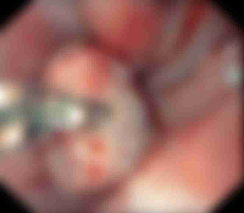
Endoscopic view of a gastric adenocarcinoma in the cardia. Source: Gastrolab/Science Photo Library
During the procedure, your doctors may find abnormal-looking areas in your stomach lining that could be cancerous. They can pass small surgical instruments through the endoscope to remove samples of this tissue for further testing (biopsy). In this manner, an upper endoscopy enables your doctors to visualize, photograph and biopsy any suspicious areas for cancer.
This imaging procedure is highly sensitive and accurate, especially when combined with endoscopic biopsy for tissue diagnosis. For this reason, doctors consider it the gold standard for diagnosing gastric cancer and it has helped increase the detection rate of early-stage cases.
Biopsy
During an endoscopy or scan, if doctors find something that looks like cancer, they will remove small pieces of this tissue for testing. This is known as a biopsy. While other tests can suggest that cancer is present, a biopsy is the only way to know for sure if an area of the body has cancer and confirm a cancer diagnosis.
Biopsies checking for gastric cancer are most frequently performed during an upper endoscopy. If doctors find any abnormal-looking areas in your stomach, they can pass instruments through the endoscope to retrieve a tissue sample. Doctors can also take biopsies from areas where the cancer has possibly spread, such as nearby lymph nodes.
Some cancers growing in deeper layers of the stomach wall can be difficult to biopsy with a standard endoscopy. If doctors suspect the cancer is located deeper in the stomach wall, they can use a different imaging test called an endoscopic ultrasound (EUS). This procedure uses a thin, hollow needle that is guided to a specific area deep in the stomach wall to take the biopsy sample.
Biopsy samples are sent to a laboratory, where a pathologist examines them under a microscope to see if they contain cancer cells. If they do, the pathologist can also determine what type of gastric cancer and how aggressive it is.
Barium X-rays
Also known as an upper GI series or barium swallow, this non-invasive test uses X-ray imaging to examine the esophagus (food tube), stomach and duodenum (first part of the small intestine). For this test, you drink a white chalky solution that contains a substance called barium. Subsequently, a series of x-ray pictures are taken.
Barium is a silver-white metallic compound that coats the lining of the esophagus, stomach and small intestine as it moves down your upper GI tract. Because X-rays cannot pass through the coating of barium, it acts as a contrast medium that improves the visualization of tumors and other abnormalities in the lining of these organs.
Unfortunately, the sensitivity of barium X-rays is low and false-negatives can occur in up to 50% of cases. In fact, the sensitivity of barium studies can be as low as 14% for early-stage gastric cancers. With the widespread availability of endoscopies and other imaging scans, barium X-rays have diminished in popularity as a diagnostic technique for gastric cancer.