Tests and Scans for Staging Stomach Cancers

Adapted from: Shutterstock
Receiving a stomach (gastric) cancer diagnosis can be shocking and unsettling. You may be experiencing all sorts of emotions and everything can seem overwhelming. While processing your diagnosis and emotions, your doctors may be acting fast and requiring further scans and tests from you. This can make the whole experience even more overwhelming. However, these tests are crucial to learn more about your cancer’s stage. This will determine the treatment options that would work most effectively for you. Ultimately, these procedures will help you and your cancer care team plan for your cancer journey ahead. Understanding how these procedures work may create a better outcome that can also positively affect your overall well-being as you begin this journey.
What is cancer staging?
Staging is the process of describing where a cancer is located and how far it has spread to other parts of the body, if at all. This determines the severity and extent of your cancer, which helps doctors predict the course of your condition (prognosis) and decide on the best treatment options for you. For instance, early-stage gastric cancer may be best treated with surgery, while advanced-stage gastric cancer would likely require treatments, such as chemotherapy, that reach all parts of the body.
How do doctors determine your cancer’s stage?

Source: Peakstock/Science Photo Library
After confirming a gastric cancer diagnosis, your doctors will then need to determine the cancer’s spread and ascertain its stage. To do this, they use different types of tests and imaging scans.
Endoscopic ultrasound (EUS)
An ultrasound is an imaging scan that uses sound waves to create a picture of your internal organs. An ultrasound image of your stomach can show the size of the cancer, how deeply it has grown into the lining of your stomach wall and/or whether it has spread to nearby lymph nodes, tissues and organs.
An EUS (or endosonography) uses a thin, lighted, flexible tube called an endoscope, which has a small camera and ultrasound probe placed at its tip. This instrument is passed through your mouth, down your esophagus and into your stomach. When the probe is put up against the stomach wall (where the cancer is located), high-energy sound waves bounce off the internal tissues and create echoes. These are converted into highly detailed images that are projected onto a video screen. Your doctors use these images to see the layers of the stomach wall and nearby structures.
If the ultrasound reveals that nearby lymph nodes are enlarged, your doctors may want to check them for signs of cancer. To do this, they can use the ultrasound images to guide a needle to collect tissue from the lymph nodes. This is known as an EUS-guided needle biopsy. The biopsy sample is subsequently examined in a laboratory for the presence of cancer cells.
Computed tomography (CT) scan

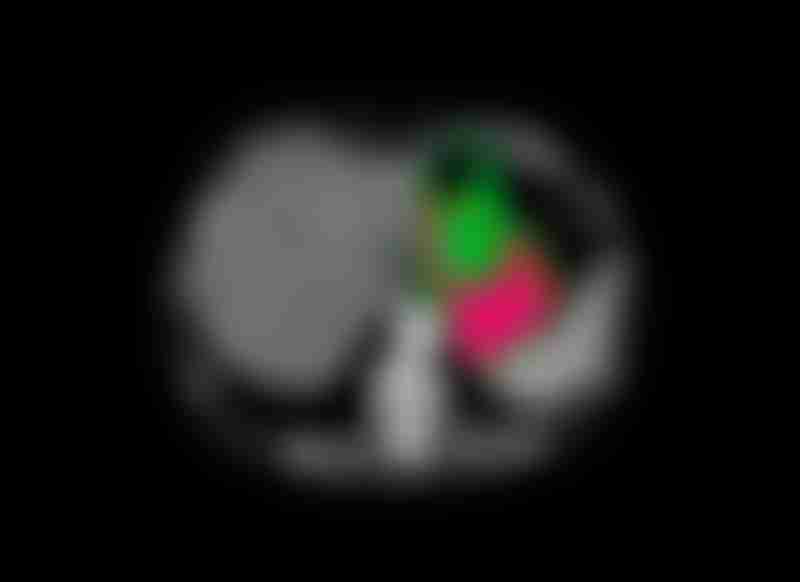
CT scan of a patient's abdomen, showing an adenocarcinoma in the stomach (highlighted in red). Source: Centre Jean Perrin, Ism/Science Photo Library
A CT scan is an imaging technique that uses x-rays to create highly detailed, cross-sectional images of the soft tissues in your body. CT scans of your chest, abdomen and pelvis can show the location of the gastric cancer and whether it has spread to nearby lymph nodes and/or other organs, such as the lungs or liver.
For this test, an x-ray machine scans your body in 360 degrees and captures pictures of your internal tissues and organs from different angles. A computer, which is linked to the x-ray machine, combines these pictures into a series of detailed, three-dimensional (3D) images that show any abnormalities or tumors. Sometimes, before the scan, you may be given a special dye called a contrast medium, which allows for more detailed visualization. This dye can be administered as a liquid to swallow or an injection into your veins.
If your doctors spot areas of suspected cancer spread during the procedure, they can use CT scans to guide a biopsy needle through your skin and toward the tumor. This is called a CT-guided needle biopsy. Once the tissue sample is collected, it can be sent to a laboratory for further testing.
Positron emission tomography (PET) or PET-CT scan
Another test used by doctors is a PET scan. This imaging technique is used to determine how far the cancer has spread to nearby lymph nodes and other organs.
For this test, a slightly radioactive sugar substance is injected into your body. This sugar substance is absorbed by cells that use large amounts of energy. Tumorigenic processes such as unregulated cell proliferation and survival require a lot of energy to maintain. For this reason, cancer cells need more energy than normal healthy cells. To meet their increased energy demands, cancer cells are likely to take up more of the radioactive sugar substance that is injected into your body. However, please be rest assured that the levels of radiation in the substance are low and not harmful.
Subsequently, a scanner will detect the presence of the radioactive substance in your body to produce images of your internal tissues and organs. The higher uptake of the substance by cancer cells makes cancerous regions of the body show up as bright hot spots on the scan.
Magnetic resonance imaging (MRI)
An MRI is similar to a CT scan, but instead of x-rays, it utilizes magnetic fields to produce highly detailed images of your internal organs and tissues. It is generally used to measure the size of tumors. Before the scan, you may be given a special dye called a contrast medium, allowing more detailed visualization. This dye is typically administered as an injection into your veins.
This imaging test is not used as often when it comes to preoperative staging for gastric cancer. Movement in the form of gastric peristalsis can result in noisy and blurry images being produced. This means your doctors may have difficulty in accurately assessing the cancer’s spread and stage using these reduced quality MR images. However, an MRI may be helpful in looking for tumors that have spread to the liver.
Laparoscopy
If imaging tests like CT or PET scans haven’t provided enough information in terms of the cancer’s spread to other parts of the body, your doctors may perform a laparoscopy. This minimally-invasive surgical procedure can help to confirm if cancer growth is limited to the stomach, or if it has spread to the liver or peritoneum. A CT or PET scan might not be able to detect cancer spread in these areas.
You will first be put under general anesthesia, so you’ll be unconscious and won’t feel any pain. Subsequently, your doctors insert a laparoscope, which is a thin, lighted flexible tube with a small video camera at the tip, through a small cut made in your belly. Highly detailed images from the camera are projected onto a video screen, allowing your doctors to observe closely the surfaces of your bodily organs and nearby lymph nodes in the abdomen. Your doctors can also remove small samples of tissue to test them for cancer (biopsy). If there’s no visible signs of cancer spread, your doctors may wash the abdomen with saline liquid, which is called peritoneal washing. The fluid is then collected and examined for the presence of any cancer cells. In some instances, laparoscopy can be combined with an ultrasound to produce better quality images.
The thought of undergoing these tests may add to the stress that you are already feeling. However, it’s important to understand the purpose of these tests and how they can help you prepare for the road ahead. By being informed, you can prepare yourself to handle any potential challenges or discomforts that may arise during treatment. This information will also enable you to have more direct conversations with your cancer care team about any concerns. By being well-prepared, you can work more effectively with them. This collaborative nature and open communication will help make your treatment journey more manageable, both for you and your doctors.