Shadow on Lung: Should You Worry?

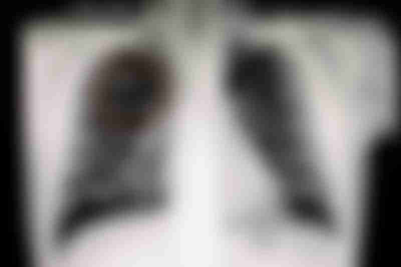
Shadow on lung seen on X-ray. Source: Shutterstock
What does a shadow on the lung mean?
A "shadow" on the lung may appear as a dense area on a chest X-ray, or as a white "spot". These are usually lung nodules, which can be found in up to half of adults during an X-ray or CT scan.
By definition, a lung nodule is smaller than 30 mm (about 1½ inches). A lung mass larger than 30 mm is usually assumed as a cancerous tumor. To establish a diagnosis, your doctor will order a computed tomography (CT) scan and other diagnostic tests.
Can a shadow on the lung be nothing?
A shadow on the lung may sometimes be nothing to worry about. While most lung nodules are non-cancerous, some may suggest the early onset of lung cancer.
How likely is a shadow on the lung cancer?
A shadow on the lung likely isn't lung cancer. According to the American Thoracic Society, less than 5% of detected nodules turn out to be cancer. A cancerous nodule is more likely in people who
- Have a larger nodule (≥30 mm)
- Are older (age ≥65)
- Smoke, or have smoked previously
- Have other risk factors for lung cancer
The risk of lung nodules becoming cancerous increases with the size of the nodule:
Size of nodule | Cancer risk |
|---|---|
≤2 mm | Extremely low risk |
2-6 mm | 0-1 % |
6-8 mm | 0.5-2 % |
≥8 mm | Nearly 10% |
≥10 mm | About 15% |
≥30 mm | Assumed as a cancerous tumor; more diagnostic tests are needed to confirm |
Data source: Larici et. al. (2017)
Even if you are at high risk for lung cancer, small nodules are probably not cancerous.
How serious is a shadow on the lung?
In most cases, finding a shadow on the lung is not serious. Benign (non-cancerous) lung nodules are commonly caused by scarring from injury (exposure to environmental irritants) or healing from an infection. Inflammatory conditions like rheumatoid arthritis can also lead to the formation of non-cancerous nodules.
If a doctor finds a nodule on a chest X-ray, they usually perform a CT scan and other tests to diagnose the condition. However, a biopsy of the nodule is needed for a conclusive diagnosis.
To avoid unnecessary stress and costs for patients, doctors will first estimate the likelihood of malignancy. This is done by looking at the nodule size and other risk factors. These include the patient's age, smoking history, and family history and more.
If the nodule has the appearance or characteristics of a benign lesion, the doctor may take a ‘wait and watch’ approach. A repeat CT scan may be ordered after some time to monitor if the nodule is growing.
A single, solid nodule that is stable (not growing) for two years is likely to be benign and there is no need for further workup.
What about suspicious nodules?
When cancer is suspected, your doctor may recommend active surveillance. You will get another CT scan in up to 12 months to check for any growth or development in the nodule. An increase in nodule diameter of >2 mm is considered growth.
Even if the nodule is subsequently confirmed as lung cancer, there is still a good chance that you can be cured. An incidental malignant nodule (<30 mm) is early-stage lung cancer. It can be treated easily and the overall survival rate is as high as 80%.
Other imaging tests and biopsy
Along with chest CT scan, a positron emission tomography (PET) scan can help determine the likelihood of a nodule larger than 8 mm to be cancer. A PET/CT scan involves the injection of radioactive sugar into the bloodstream. A rapidly growing and cancerous tumor absorbs more sugar compared to normal cells. The radioactivity in malignant nodules produces a higher contrast under a CT scan.
- For healthy individuals at high risk, a surgical biopsy is the gold standard for nodules. Visual inspection and pathologic analysis of nodules and neighboring lymph nodes can be done during the operation. If the nodule is indeed cancerous, it can be resected immediately. If the lymph node is affected, the surgery can be converted to a lobectomy (or segmentectomy).
For a nonsurgical approach, sampling of nodules can be done via
- the airway (bronchoscopic biopsy) or
- the chest way using a needle (transthoracic needle biopsy)
The pathologists assess the nodule specimen to make a diagnosis. A treatment plan is then discussed and determined between the doctor and the patient.