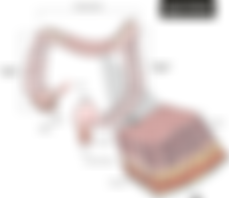
Anatomy of the Colon

Source: Shutterstock.
The large intestine, an organ that is about 5 feet (150 cm) long, consists of four sections.

Anatomy of the colon, also known as the large intestine.
Adapted from: Shutterstock.
The first section known as the ascending colon begins with a pouch known as the cecum where undigested food matter comes in from the small intestine. It is located towards the right side of the abdomen and continues upwards towards the transverse colon.
The transverse colon is the second section that goes across the abdomen from right to left.
Together, the ascending and transverse colon make up the proximal colon.
The third section, called the descending colon, travels down the left side of the abdomen.
The fourth, S-shaped section known as the sigmoid colon is connected to the rectum leading to the anus.
The descending and sigmoid colon are collectively called the distal colon.
The rectum is located immediately after the sigmoid colon at the lower part of the large intestine. It is approximately 15 cm long, and stores waste received from the colon that will be passed out through the anus.
Layers and cells found in the colon and rectum

Cross section of the large intestine lining.
Source: Shutterstock.
The large intestine is a muscular tube that absorbs water and nutrients, and helps move digested food matter to the rectum.
Different cells, tissues and muscles found in the colon help it to perform its different functions of moving digested food, absorbing water and nutrients, and also secreting hormones to regulate appetite.
Similar to the colon, the rectum is also made of different layers of cells and tissue.

Cross section of the rectum.
Source: ResearchGate.
Layers of the colon and rectum
A cross section of the colon can be divided into the following layers:
- Mucosa: A layer where cells that secrete mucus, produce hormones and absorb water and nutrients are found.
- Submucosa: A layer of fibrous tissue found beneath the mucosa.
- Muscularis propria: A layer consisting of circular smooth muscle and longitudinal smooth muscle important for peristalsis.
- Subserosa and serosa: A layer of connective tissue that forms the outer lining of the colon. The serosa is only present in the ascending, descending and sigmoid colon.
Cells in the mucosa
Enterocytes are the large intestine’s absorptive cells that take up water and nutrients from the digested food. Microvilli found on the surface of these cells help to increase surface area for more efficient absorption.
Goblet cells are found in the mucosal layer of the large intestine’s inner lining. These cells are responsible for secreting mucus to protect the lining from infection and inflammation. The mucus also helps to maintain a healthy environment and relationship with gut bacteria, which play crucial roles in digestion and managing appetite.
Enteroendocrine cells that are also found in the mucosal layer are responsible for secreting hormones. These hormones include PYY and GLP-1, which are responsible for reducing appetite and inducing satiety, and promoting insulin secretion respectively.
Cells in the muscular layer
In order to move food from one end of the colon to another, different soft muscle tissues are required. The muscular layer consists of two layers of smooth muscle; the inner, circular layer, and the outer, longitudinal layer. These muscles contract and relax in a process known as peristalsis, a wave-like motion that helps move the food along the large intestine.